![]()
|
|
|
At Great Danes4U, (a Great Dane Puppy Breeder in the Springfield, Missouri - Marshfield, Missouri 65706 has Quality Great Dane Puppies for Sale. Your
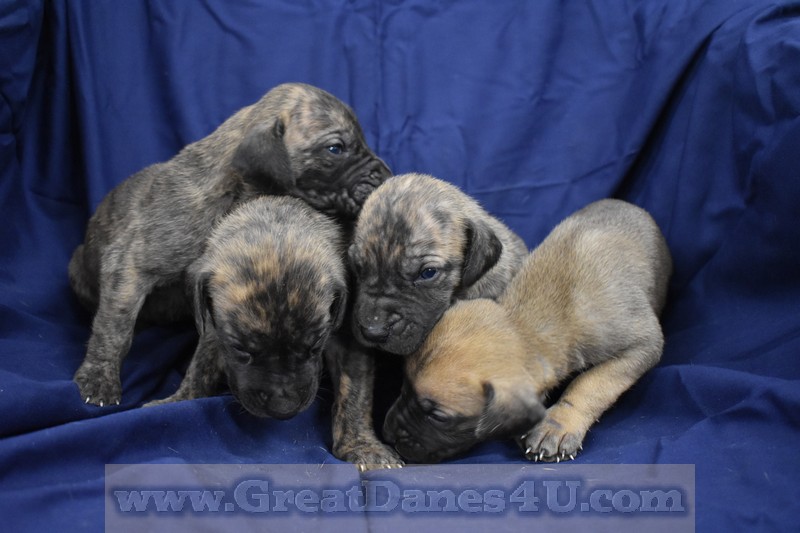
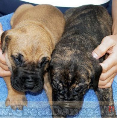
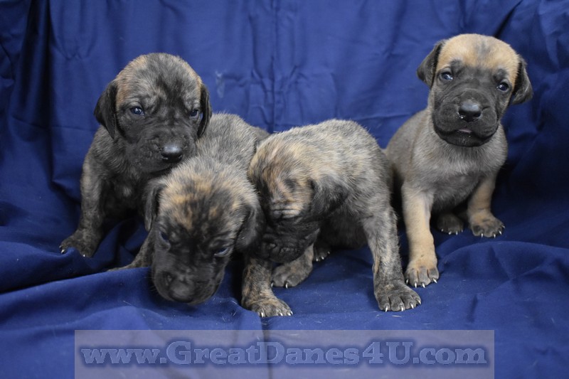
Great Dane puppy could be a Fawn Great Dane puppy, or a Brindle Great Dane puppy. Most of our Fawn & Brindle Great Dane stock is +5-Generation Color-Pure AKC Registered. All are Micro chipped plus all
older parents and Grand Parents are DNA Profiled. Most of our stock are OFA Certified, (Orthopedic Foundation for Animals), if age appropriate. Enjoy Free Information about Great Danes Appearance, Standards, K-9 Care,
this Breeder's Policies, Dane History and many Pictures of Great Danes on this
FREE Website. GreatDanes4U uses this site to promote Healthy, Trained, Socialized Multi-Generation Color-Pure FAWN and BRINDLE AKC Great Dane Puppies. Many Great Dane puppies are Breeding Quality and there are Show Potential pups available as well. Stud service also available. If you are using a "cell phone" to access this site try: www.GreatDane4U.com *It is a static site but easier for cell phone access. |
|||
|
"TIGERS" |
 |
 |
 |
|
Please: fly in, and Drive a Rental vehicle home, or drive both ways.
**Recently "1-855-PetList" picked up a pup for a delivery!** Air shipping includes Crate & crate prep, Vet visit, Health Certificate, Air Fare, (based on size of crate & weight + puppy weight & logistics to make it happen: *Pups under 35# add $500, Between 35 & 75#'s add $600. Air S&H Prices are only for departures from: Springfield, MO. Airport, (SGF). Note: Out of area, (3 hours away) air shipping Add $200. **If using a
Cell phone try a static or stale site for cell phones:
www.GreatDane4U.com It does not get
updated. This site does get updates, but you need a PC.** |
|||
|
Use Blue Drop-Down, (Cascading) Menu Bar.
***Click on Thumbnails Below to enlarge images.*** |
|||